Cultural heritage science—sometimes referred to as conservation science—is the scientific study of the materials associated with art and archaeology, including objects that can be moved and objects that must be studied on-site.
Cultural heritage scientists study a wide range of materials that can include metals and corrosion products; textiles and dyes; mineral and organic pigments; gems; composite biomaterials, such as leather, vellum, parchment, and wood; paper and papyrus; natural and synthetic polymers; and even biofilms and biodeterioration. Perhaps the materials of greatest interest to the American Ceramic Society community would include fired ceramics, glazes, adobe, enamels, mortar, glasses, and ceramic nanocoatings.
Cultural heritage scientists apply their unique skills in art museums, universities, and regional or national centers for the conservation of art or to landmarks, such as archaeological sites or architectural buildings. This highly interdisciplinary profession requires expertise in physical sciences (chemistry, physics, biology, geology, and material science), forensics, humanities (art history, archaeology, archaeometry, art conservation, ethics, and history), engineering, computer science, and economics. Therefore, collaborations are critical to success, and particularly strong working relationships exist between conservators and curators. Within ACerS, the recently established Art, Archaeology, and Conservation Science Division supports the cultural heritage science community.
Cultural heritage scientists assume various roles depending on the circumstances surrounding an artwork or a particular issue—a doctor one day, a detective the next, or even an explorer the following day. For example, if crystals spontaneously appear on a ceramic artwork, museum curators may ask scientists: “What is it? Where did it come from? Why is this happening? How can this be prevented?” Alternatively, scientists may be asked to address questions of authenticity. If an art museum plans to acquire an object, a scientist might be asked to confirm that the materials used for the object’s fabrication are consistent with the date of attribution. Sometimes, a scientist is asked to reverse engineer a work of art to determine how it was made or what technology was used to make it.
Regardless of the question that sparks an investigation, the scientist faces unique challenges when working with art. Sampling a work of art is very limited because artwork is precious and should not be consumed—or visually impacted—during an analytical investigation. Therefore, scientists use nondestructive analytical tools first. These include visual examination, usually with a stereomicroscope, and possibly various forms of radiation that could include ultraviolet (UV) or infrared (IR). Computed tomography (CT), X-radiography, and elemental identification using X-ray fluorescence (XRF) also are tools of cultural heritage science that can be applied without sampling. When sampling a work of art is deemed appropriate, scientists routinely use scanning electron microscopy–energy dispersive spectroscopy (SEM-EDS), X-ray diffraction (XRD), and Fourier transform infrared spectroscopy (FTIR). This article will highlight some of the specialized characterization tools used by cultural heritage scientists, using as examples analytical investigations of artwork from the Walters Art Museum in Baltimore, Md.
Revealing tool marks with stereomicroscopy
The ultimate sleuth Sherlock Holmes always has a magnifying glass ready. Similarly, a cultural heritage sleuth obtains a close look as the preliminary step for any examination of a work of art, and an optical stereomicroscope can enhance such an examination. For example, tool marks from abrasives on carved jade from the Americas provide evidence that helps distinguish between ancient and modern techniques and can inform questions of authenticity. Ancient American societies carved jade extensively using loose abrasives that created irregularly and variably sized marks. Modern cottage industries also supply collectors with carved jade objects that imitate ancient production, although these techniques make use of fixed abrasives that leave marks appearing as fine, regularly spaced parallel lines.1
During a recent study of a jade artifact (Figure 1), surface tool marks and silicone molds of the surface were inspected using several imaging methods, including stereomicroscopy, SEM, and reflectance transformation imaging (RTI). RTI, once referred to as polynomial texture mapping, is a photo-documentation technique that uses computer algorithms to capture micrometer-scale detail, texture, and even color of surfaces.2 This study confirmed authenticity of the artifact and showed that lower-cost RTI provides comparable results to traditional, but expensive, SEM imaging.
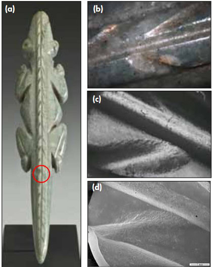
Figure 1. Authentically ancient or not? (a) Jade Crocodile Effigy Pendant (WAM No. 2009.20.273) (15.7 cm high) carved in Atlantic Watershed-style and attributed to ancient Costa Rica; gift of John G. Bourne Foundation, 2013. (b) Toolmarks are difficult to discern on Crocodile’s surface in this 10-mm-long photomicrograph. (c) RTI and (d) SEM of silicone impression from Crocodile at 40x clearly document a surface prepared using loose abrasives. Credit: Susan Tobin; WAM
Illuminating art with UV and IR
Various types of illumination are central to the toolkit used to investigate art. Generally considered nondestructive during the short exposure times of an examination, UV and IR can reveal or differentiate many materials that the unaided eye cannot. For example, Figure 2 illustrates an inlaid steatite (or “soapstone”) Egyptian Pectoral with Scarab from 1292–1070 B.C. The pigment known as “Egyptian blue” is a synthetic calcium copper silicate compound with a characteristic bright-white visible-light-induced IR fluorescence.3 Egyptian blue is one of few artists’ pigments with an end-of-use date. Although Egyptians used it from at least 2500 B.C., the method for making it appears to have been lost around the fall of the Roman Empire, A.D. 400. Therefore, the presence of Egyptian blue pigment indicates a paint created in ancient times, as opposed to a more modern restoration that might have been applied to enhance the value of an object for sale.
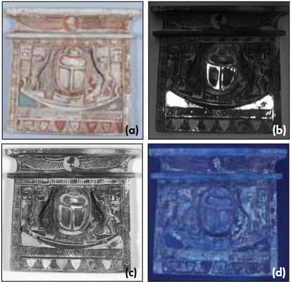
Figure 2. (a) Under normal light, this steatite Egyptian Pectoral with Scarab (WAM No. 42.91) (9.5 cm high) from 1292–1070 B.C. shows many colored inlays. (b) Using visible-light-induced IR fluorescence, Egyptian blue pigment appears bright white. (c) Reflected IR easily distinguishes Egyptian faience (white) from the steatite (gunmetal gray). (d) Remnants of glaze, which are preserved primarily in the recesses and interstices of incised lines, appear bright-white under UV. Credit: Susan Tobin; WAM
At the lower edge of the pectoral, the register of stylized lotus blossoms retains four of the original shield-shaped inlays that appear indistinguishable from the steatite support under normal light (Figure 2(a)). Vsible-light-induced IR fluorescence (Figure 2(b)) indicated that these regions contained Egyptian blue pigment. However, reflected IR (Figure 2(c)) clearly showed that these stylized lotus blossoms are made from a different material that is not steatite, but probably Egyptian faience—a partially sintered quartz-containing ceramic with surface vitrification. Under UV radiation (Figure 2(d)), the fluorescent remnants of a glaze on top of the steatite were clearly visible in the recesses and interstices of the incised lines and suggested the steatite was fired, probably to harden the normally soft stone substrate and set the glaze.
Peering inside—CT scanning and X-radiography
Examining an artwork’s interior often is instructive for determining how an object was made, but it is very difficult or sometimes impossible to achieve. X-radiography can help discover how an object was made with details regarding its construction or use. For example, it can reveal joins or points of attachment that are not visible to the eye. It also can show the contents of internal cavities of hollow objects, which provide clues to how the civilization used them.
An X-radiograph records variations in density, with greater densities appearing lighter because of absorption of X-rays. Although it is an incredibly useful tool for many artifacts, interpreting an X-radiograph can be quite complicated, because it records the density variations of a three-dimensional object onto a two-dimensional surface (formerly film, but more commonly today a digital plate). CT scanning compiles X-ray images obtained in thin sub-millimeter- or even micrometer-thick slices. As a result, CT scan images may be easier to interpret. In the case of the 2,000-year-old ceramic figure from ancient West Mexico (Figure 3), X-radiographs suggest that the head, body, and legs were hand-built, but it was difficult to clearly discern just how. A CT scan allowed a clear view of the interior walls, making it apparent that overlapping slabs of clay were used to create the figure. Additionally, it appears that the legs, torso, and head were created separately and then joined.4

Figure 3. (a) Standing Female Figure (WAM No. 2009.20.62) (38.5 cm high) from Nayarit in ancient West Mexico (Lagunillas “C” type) was produced between 300 B.C. and A.D. 200 from burnished, slip-painted earthenware; gift of John Bourne, 2009. (b) X-radiograph and (c) CT scan of the figure show joins, suggesting that sections were created separately then affixed to one another. Credit: U. of Maryland Medical Center
Identifying chemical elements—XRF
Some consider XRF the most powerful nondestructive analytical tool in the arsenal of the cultural heritage scientist. During the past 50 years, XRF has helped build a rich database of the elemental compositions of artwork surfaces. In very dense materials, such as gold-based materials, XRF can identify elements to a depth of 25 μm below the surface. In less dense materials, XRF can identify elements several hundred micrometers deep—sometimes at parts-per-million concentrations. Historically, XRF instrumentation used for art analysis was comparatively bulky or had to be used in a lead-lined room, which required transportation of artwork to the analytical instrument. This is not an issue when the object is small and portable, such as the Tiffany Iris Corsage Ornament pictured in Figure 4.
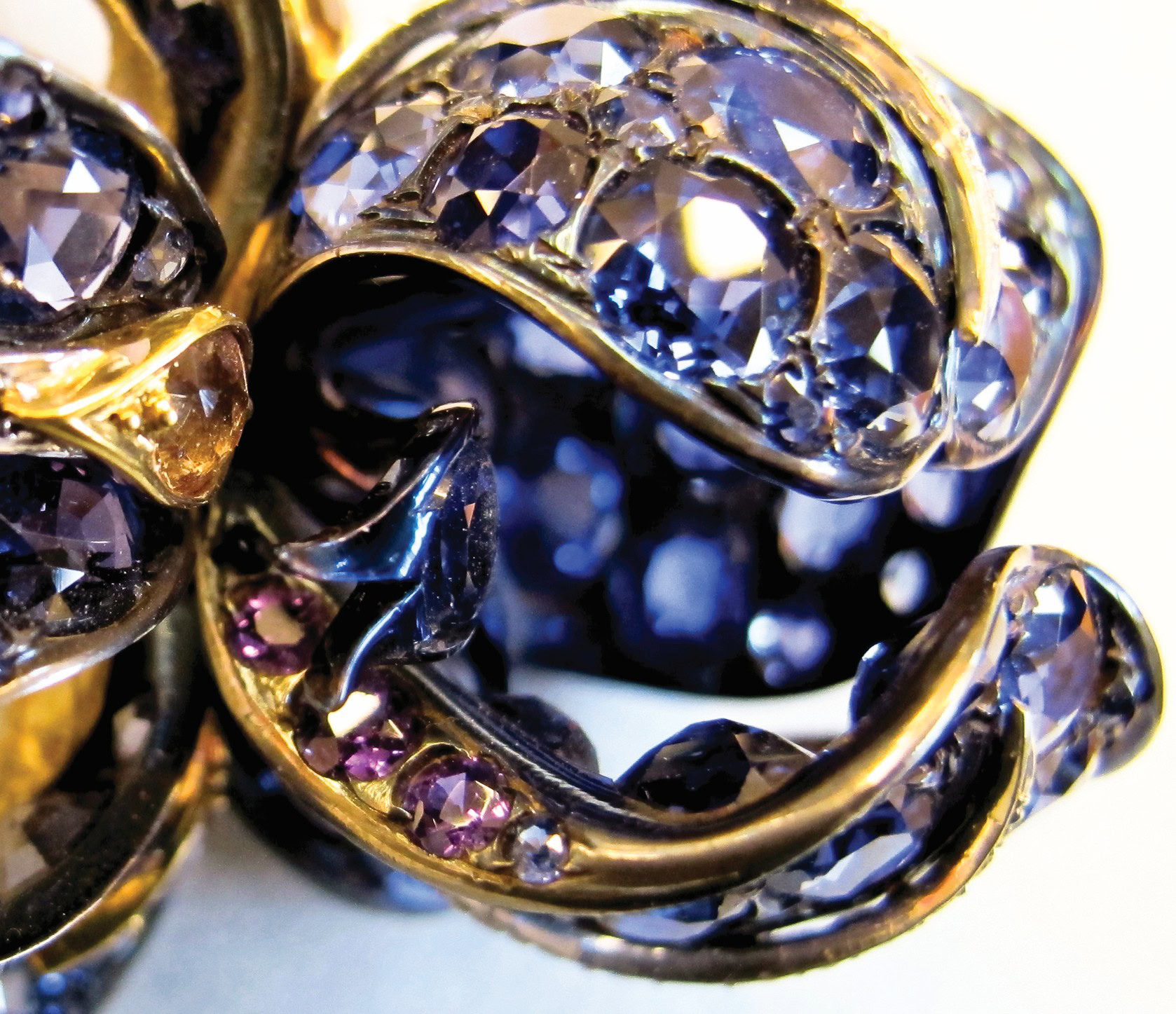
Figure 4. XRF analyzes elemental differences between blue and violet Montana (or Yogo Gulch) sapphires used to form the petals of the Iris Corsage Ornament. Credit: Susan Tobin; WAM
Many objects worth studying are not so easily moved. During the past decade, portable XRF instrumentation has come of age to allow routine, on-site analysis of monumental artifacts that cannot be moved. For example, certain sections of the life-sized, wall-mounted Adam and Eve glazed terracotta relief shown in Figure 5 were suspected of being restorations (Figure 5(c)). Using a portable instrument, XRF analysis distinguished original Renaissance parts from later restoration additions based on elemental differences in the glazed surfaces.5
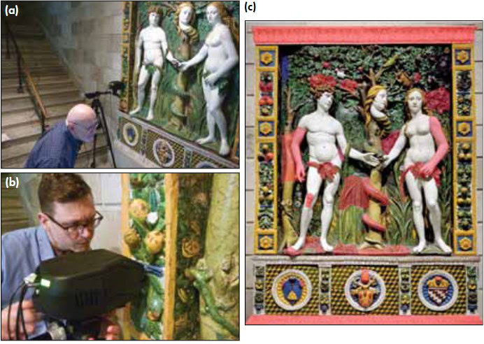
Figure 5. (a) and (b) A portable XRF spectrometer analyzed the glazed surface of the life-sized Adam and Eve (WAM No. 27.219) (280 cm high) created by Giovanni della Robbia in Italy circa 1515. (c) The red shadowing indicates areas of suspected restorations, which were confirmed by XRF. Credit: Susan Tobin; WAM
Discovering materials interactions with FTIR
FTIR can be extremely useful to identify materials by providing information regarding how elements are combined. In the instance of the ceramic Mycenaean Jug in Figure 6(a), FTIR helped answer important questions about the appearance of a crystalline growth on the interior rim of this low-fired ceramic vessel (Figure 6(b)). The crystals appeared while the object was on display, and museum conservators knew the crystals were not part of the jug’s archaeological history. Indeed, the crystals actually were forcing their way through the ancient slip-decorated surface, causing damage to the object, as observed with an optical microscope (Figure 6(c)).
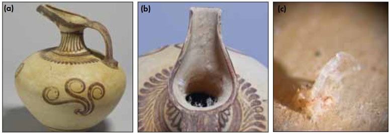
Figure 6. (a) Long-Beaked Jug (WAM No. 48.2098) (27 cm high) from Mycenae, circa 1425 B.C.; museum purchase in 1957. (b) Top view shows growth of new, unknown crystals in jug opening. (c) This microscopic image of the 0.5-mm-wide crystals in-situ shows deterioration of the ceramic body from volume expansion during crystallization of hydrated calcium acetate. Credit: Susan Tobin; WAM
The spectrum of an FTIR analysis conducted through a microscope in transmission mode, with a microsample compressed between diamond plates, identified the crystals as a hydrated calcium acetate compound.6 Organic acids in the form of acetates and formates from exhibition materials can be a serious pollution problem in the museum environment. Potential polluting organic materials include wood (particularly oak) and wood products, fabrics, and adhesives. In this case, the analytical results led to a federal grant through the Institute of Museum and Library Services to retrofit exhibition cases. This included replacing fabrics in cases with paint to diminish exposure of objects to pollutants. This project resulted in a change of protocol and materials for all new case construction. In this way, materials studies informed materials selection decisions regarding exhibit construction to best preserve collections of cultural heritage for the future.
Deterioration or artistic design—XRD and SEM
Determining if the appearance of an artwork results from its history and merits preservation or if its appearance is a product of decomposition or deterioration is not always as obvious as in the preceding case. For example, conservators noticed the presence of a white crystalline material on the surface of the Venetian glass Ewer (Figure 7(a) and (b)) while preparing it for exhibition. Because this artwork was created in the late 19th or early 20th century with the intention of appearing ancient or antique, the question arose whether the accretion indicated an unstable glass composition or if it was evidence of the “antiquing” process used by the creator and, therefore, ought to be preserved as part of the object’s history.
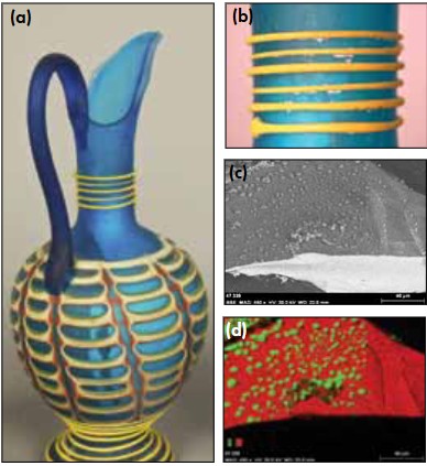
Figure 7. (a) Italian glass Ewer (WAM No. 47.339) (17.6 cm high) by Salviati and Co., produced in the late 19th or early 20th century to mimic the style and appearance of ancient glass. (b) Neck detail shows white encrustation on the glass. (c) Backscattered electron image of a sample from the surface shows the polygon shape of surface encrustations. (d) Energy-dispersive map of fluorine (green) and silicon (red) shows fluorine located in the encrustation but not in the underlying glass. Credit: Susan Tobin; WAM
XRD analysis identified the accretion as malladrite, a sodium fluorosilicate. This mineral occurs in the vicinity of Italian volcanoes, including Mount Vesuvius, near where the object was created in Venice. However, the question remained whether the object itself was decomposing. Extracting a small sample that included the glass and the accretion for SEM-EDS analysis showed with certainty that the white material had been applied, because fluorine was detected only in the accretion, not in the underlying glass substrate (Figure 7(c) and (d)). In this case, material analysis informed the proper conservation of the object during its preparation for exhibition—the accretion was evidence of the maker’s antiquing technique, thus it was preserved.7
Teasing out provenance with neutron activation analysis
Sometimes the scientific study of artwork materials helps inform the history or origin of an unprovenanced, undocumented artwork. Such was the case for a vast collection of more than 1,000 Byzantine ceramic tiles and tile fragments that were purchased as a group by the Walters in 1956 from an art dealer. This dealer purchased the collection at the Istanbul Bazaar, so little information existed about the context or geographical origin of this important collection of figurative and decorative tiles. Made approximately 1,000 years ago, such tiles are rare and likely served as decorative schemes for the interior of small chapels or sacred spaces.
A similar group of tiles of known provenance in the collection at Dumbarton Oaks (Washington, D.C.) was excavated by M. Ramazanoglu from a site in Constantinople, the Byzantine capital known today as Istanbul, Turkey. The Walters tiles appeared to be similar but differed in significant ways. Therefore, scientists compared trace element compositions of the ceramic bodies to try to establish a secure connection to the Byzantine capital. Drilled samples from the ceramic body of the Walters tile fragments were compared with Dumbarton tile fragments using neutron activation analysis (Figure 8).
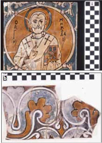
Figure 8. (a) Painted icon wall tile of St. Nicholas (WAM No. 48.2086) (15 cm high) from Byzantium, 10th century; museum purchase in 1956 and partial gift of Robert E. Hecht Jr., 1957. (b) Fragment of Byzantine tile (WAM No. 48.2086 CV2) analyzed by neutron activation analysis. Credit: Susan Tobin; WAM
Cluster analyses of the neutron activation results for 53 major, minor, and trace elements at parts-per-million concentrations revealed the Walters and the Dumbarton ceramics were of the same compositional group, providing strong evidence of a substantial connection to the Byzantine capital for the Walters tiles.8
Work in-progress: Examining a pigment particle with FIB and TEM
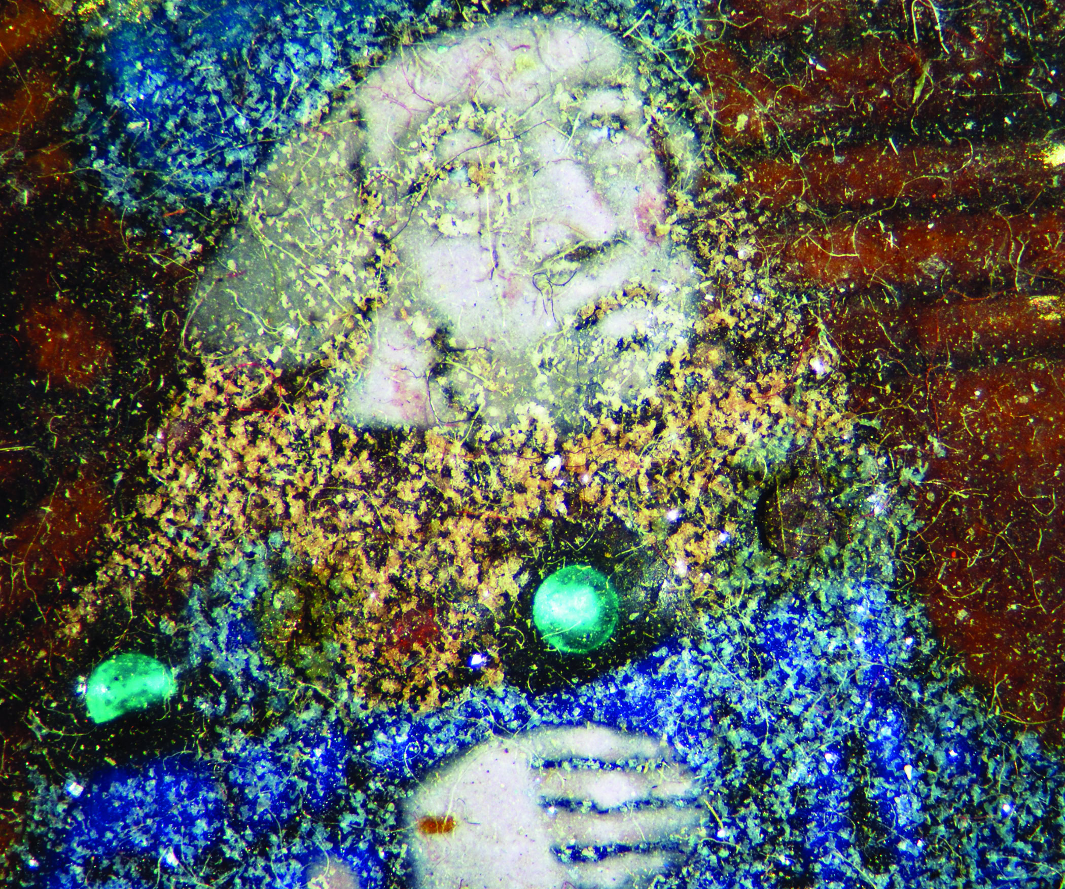
Perhaps one of the most exciting opportunities for materials analysis in cultural heritage science occurs when a conservator observes something unusual in an artwork, such as a material beyond common experience. Determining what it is, or simply knowing more about it, provides potential lessons about the artwork or its materials. This situation occurred at the Walters with the observation of unusual purple, gemlike particles embedded in paint used for the Portrait of a Bearded Man mummy portrait, produced in Egypt around A.D. 170 (Figure 9(a)). The purple paint was used for the figure’s clavi, a fashion element on the shoulder of a toga used to indicate senatorial or equestrian aristocratic rank (and from which today’s word clavicle, meaning shoulder bone, comes).
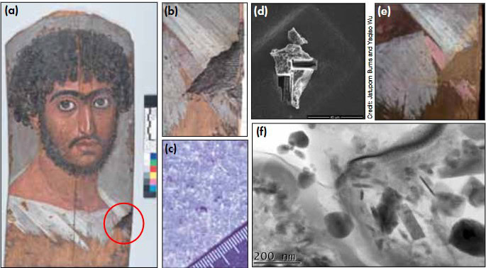
Figure 9. (a) Portrait of a Bearded Man (WAM No. 32.6) (40 cm high) is an encaustic on wood Roman-Egyptian mummy portrait circa A.D. 170. (b) Detail of lower right side of portrait showing original mummification residues on top of sparkly purple paint. (c) Detail of gemlike particles with a millimeter-scale bar. (d) Focused ion-beam slices of a purple paint particle. (e) Purple paint under UV radiation shows characteristic orange fluorescence of an organic lake pigment. (f) TEM micrograph of the pigment’s interior, including a light gray matrix, black irregularly-shaped spherical parrticles, and needle-shaped particles rich in aluminum. Credit: Jatuporn Burns and Yaqiao Wu
Elemental analysis of the unusual purple particles using XRF revealed the presence of chromium and iron. Usually, chromium indicates a modern pigment because it was not used extensively as an intentional coloring agent until after the element’s “discovery” in 1797. However, resinous residues of the original mummification process covered the purple paint, confirming that the unusual purple pigment particles were ancient, not modern. Given the gemlike appearance, it was suggested that semiprecious stones, perhaps ground-up garnets or spinels, were mixed into the paint to enhance the perceived status in the afterlife of the individual portrayed (Figures 9(b) and (c)). To identify this material, conservators removed a tiny sample of the unusual purple paint, retrieving a single particle about 20 μm in diameter for analysis—too small for XRD and susceptible to damage or burning during Raman analysis.
The single particle was mailed to Darryl Butt, distinguished material science and engineering professor at Boise State University (Idaho). His team manipulated the particle with an eyelash for SEM–EDS analysis and confirmed the presence of chromium. Subsequently, he used a focused ion beam (Figure 9(d)) to create thin sections of the particle for analysis using transmission electron microscopy (TEM). The results showed that the interior of the particle contained an organic phase rich in aluminum, potassium, and sulfur, with small amounts of iron.
Although this examination is a work in progress, these findings are consistent with the use of a lake pigment, corroborating initial findings using ultraviolet radiation that revealed the characteristic orange fluorescence of a lake pigment (Figure 9(e)). Lake pigments are created by affixing an organic dye onto an inorganic substrate, usually using a polyvalent metal ion as a mordant that joins them. Structurally, lake pigments are poorly understood. However, the TEM images from this study provide the first-ever visualization of the internal structure of an ancient lake pigment (Figure 9(f)). It remains to be determined if the chromium was intentionally added during the creation of the lake pigment, perhaps to modify the hue, or if the occurrence is unintentional. XRF analysis of a second mummy portrait also detected the presence of chromium in the purple paint, suggesting that a mordant rich in chromium was favored in ancient Egypt.
Preserving art for the future
Besides characterizing artifacts, cultural heritage scientists apply materials science knowledge to appropriately conserve artifacts for future study and appreciation. To this end, the Walters is collaborating with the University of Maryland to explore and develop atomic layer deposition (ALD) of amorphous aluminum oxide on silver art to add an imperceptible, stable barrier coating to reduce tarnish. This project is funded by the National Science Foundation.
Tarnish, or the corrosion of highly polished silver surfaces, is a monumental problem for art collections throughout the United States and the world. Removing tarnish by polishing in preparation for exhibition removes silver metal. In fact, if a hallmark stamped onto silver to a depth of 0.5 mm was cleaned every few months, the hallmark would disappear in just more than 40 years.9 The development of a long-lasting barrier coating to reduce tarnish, suitable for one-of-a-kind silver artifacts, would significantly reduce the costs and labor devoted to maintenance and preservation of silver artwork.
The standard practice in art conservation to reduce silver corrosion is to hand-apply a solvent-based polymeric coating on artwork—a tedious process. Polymer-based barrier coatings reduce the reaction of silver metal with tarnishing pollutants, but they have a lifetime of only 10 to 20 years. ALD, a technique developed by nanotechnology researchers, may prove an alternative preservation strategy. The technique is being refined to precision-deposit nanometer-thick films of metal oxide on silver-metal artifacts to reduce corrosion (Figure 10). The service life of the metal oxide coating could be more than 100 years, but by mixing getters, such as zinc oxide, into the deposited film, the lifetime of the coating may be extended by many hundreds of years.
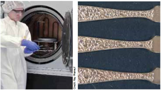
Figure 10. (a) Amy Marquardt, graduate researcher at the University of Maryland Nanocenter, prepares samples. (b) At their 2013 annual meeting, U.S. art conservators compared three 70-year-old silver art objects: one with traditional polymeric coating (top), one with the new ceramic ALD coating (middle), and one without coating (bottom). All of the 27 professional evaluators agreed the new ceramic coating was acceptable for museum exhibition. Credit: Susan Tobin; WAM
Getting involved in cultural heritage science
There are many ways to become involved in cultural heritage science. The NSF solicited proposals in 2010, 2011, and 2012 under the SCIART and CHS programs and now funds unsolicited research in the field. Increasingly, art museums feature scientific studies of art alongside more traditional stylistic or art historical presentations. If you appreciate this, make sure you let the museum know so that such programming can continue. Some major museums have science laboratories and may offer volunteer opportunities.
Of course, professional societies, such as ACerS, are always a great way to extend professional networks outside of personal areas of expertise. The AACS Division of ACerS is looking for volunteers to help with fundraising, workshop organization, and annual meetings. For people looking to get into the field, graduate and postgraduate educational opportunities in cultural heritage science continue to increase, with programs at Harvard, Northwestern, and the University of Delaware. Get involved—learn what you can bring to the study of cultural heritage and what ancient artifacts may be able to teach the modern ceramic engineer.
Transferring cultural heritage science studies to materials science today
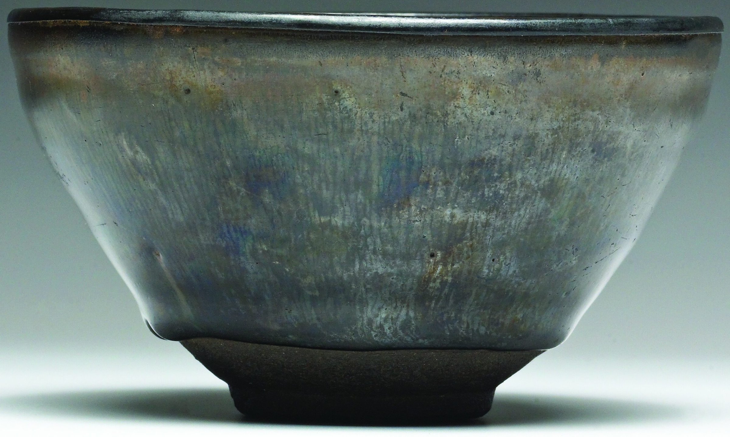
Will the study of 1,000-year-old Asian “hare’s fur” glazes yield improved magnetic materials based on epsilon-phase iron(II) oxide, ε-Fe2O3? Credit: Susan Tobin; WAM
Might studies of the deterioration of enamels produced 500 years ago in France result in improved glass for future radioactive waste storage? Credit: Susan Tobin; WAM

Can understanding the degradation mechanisms of 100-year-old cadmium sulfo-selenide pigments result in superior semiconductor performance? Credit: Susan Tobin; WAM
ACerS Art, Archaeology, and Conservation Science Division
The AACS Division mission is to advance the scientific understanding of materials found in ceramic art and to provide information that aids in the interpretation and preservation of traditional ceramic art and artifacts as well as the techniques and technologies used in their creation. AACS strives to help ACerS members better appreciate the artistic side of ceramics; work cooperatively with others in the field (e.g., craftspeople, historians, archaeologists, curators, conservators, and conservation scientists); attract and train the future workforce in this area; stimulate interest and foster interactions in this area; reconstruct older ceramic technologies; and improve public understanding and appreciation of ACerS, professional societies, and ceramists and ceramics (artistic and industrial). For more information, visit www.ceramics.org/divisions/art-division.
Walters Art Museum celebrates eight decades of art
More than two-thirds of the 35,000 objects at the Walters were acquired by the Baltimore liquor merchant and railway tycoon, William T. Walters (1819–1894), and his son, Henry (1848–1931). The elder Walters began opening his home to the public in 1874, a tradition he continued almost annually. The proceeds of these openings, where admission cost 50 cents, were donated to the Baltimore Association for the Improvement in the Condition of the Poor. Following William’s death, Henry continued to build the collection, opening his palazzo-style museum building to the public in 1909. The gallery’s buildings and contents were given to the city of Baltimore when Henry died in 1931. Now known as the Walters Art Museum, it opened as a public institution in 1934. That same year, the Walters hired a staff scientist, becoming the third art museum in the U.S. to do so, after the Fogg at Harvard and the Freer Gallery. The 80th anniversary of the Walters opening will be celebrated with a special exhibition, From Rye to Raphael: The Walters Story, on view at the museum from October 26, 2014. For more information, visit www.thewalters.org.
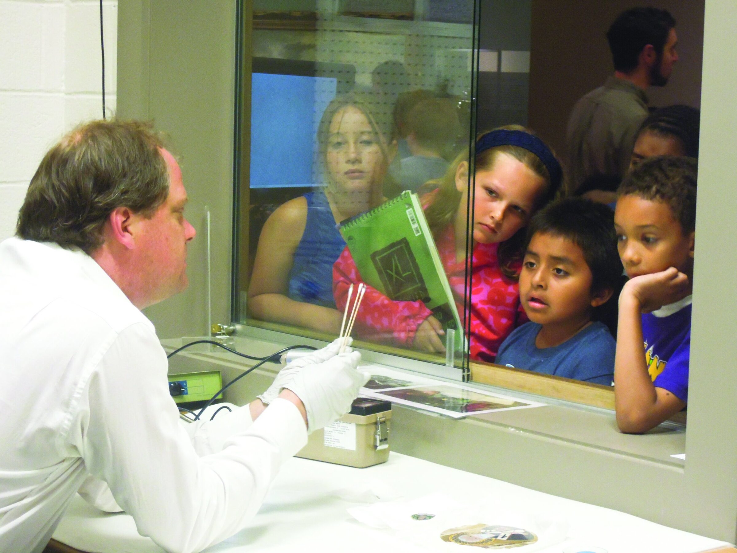
The Walters Art Museum introduces Baltimore youngsters to conservation science. Here, Glenn Gates explains how nanotechnology coatings can preserve silver art and prevent metal loss from traditional, abrasive polishing. Credit: Susan Tobin; WAM
Acknowledgments
ALD work was supported by NSF Award No. 1041803. Julie Lauffenburger and Greg Bailey at the Walters greatly helped with this article, and their assistance is much appreciated. Photography by Susan Tobin unless otherwise indicated.
Read more: “Glossary of tools for discovery in conservation science“

Related Articles

Bulletin Features
Emerging Professionals: Science for Society & Future Focus
One small tweak to the lens of materials research, one giant leap for mankind By Rishabh Kundu and Ryan C. Eaton One of the clearest existential threats facing humanity is anthropogenic climate change. The dire consequences to ecological stability and human prosperity if the status quo is maintained are thoroughly…

Bulletin Features
Emerging Professionals: Research Articles
Experiential learning: Developing the next generation of engineers By Ryan Eaton When a measure becomes a target, it ceases to be a good measure. Goodhart’s law, coined in reference to monetary policy, is readily applicable to engineering education. When students begin optimizing their study habits to pass an exam rather…

Bulletin Features
Durable and programmable metasurfaces enabled by phase change materials
Controlling light with high spatial precision enables technologies ranging from imaging and sensing to communications. Traditionally, optical components such as lenses and filters rely on bulk materials and fixed geometries, which limit their ability to adapt dynamically. Metasurfaces offer a fundamentally different approach. These materials consist of planar arrays of…